System Integration Solution

Turbo Scan自動化快速玻片影像掃瞄系統

 Automated Scanning and Imaging Control System


產品應用簡介

Solutions for Automated Microscopy and Imaging!!
Objective Imaging公司的自動化高速掃描玻片影像系統(Surveyor Automated Scanning and Imaging with Turboscan),利用相機硬體觸發同步訊號方式,同時控制自動移動平台的移動速度,可在載台高速移動的狀態下進行連續影像擷取,實現顯微光學中高倍率、高解析、大範圍的影像拼圖掃瞄整合應用 。

快速的拍照掃描整個玻片或樣品,讓樣品內的觀察目標,快速的呈現出完整樣貌,讓專業研究者可以在很短的時間內,了解整個樣品細微結構與組織形態,解脫顯微鏡高倍圍觀察的視野侷限,適用病理組織切片、金相組織大範圍掃瞄和半導體晶圓大範圍掃瞄拼圖等應用需求。

TurboScan只需簡單的滑鼠點擊設定,便可以檢測任何想要觀察或確認的玻片位置,對於研究者後續重覆檢測確認、研討、發佈等工作,都可以節省很多的時間。

利基於OASIS Automation Controller與特殊的相機觸發功能,TurboScan提供一套理想的自動化掃片解決方案,包含樣品的定位、掃描、隨機拍照、自動化拍照存檔等等。

結合Prior PL200玻片自動載入機,可容納兩百片玻片,更可以達成大量的自動化玻片掃描工作,適用於大量病理切片顯微影像掃描應用,對於有大量玻片顯微拍照的工作者,是最佳的選擇。

相關應用:玻片掃描、病理切片掃描


功能特點:

Mosaic Acquisition

  • Turboscan for fast continuous scan and acquire

  • Standard scan for step and acquire for low light applications

  • Snake and raster scanning

  • User-defined settle time at each field

  • Pause and continue scanning mode

  • Grab individual fields into mosaic with key-press

  • Acquire at full or reduced resolution

  • JPEG compression option

  • Image streaming for mosaic sizes limited only by free disk space

Specimen Map

  • Stage and pattern views

  • Point and click relocation to position or pattern field

  • Real-time graphic display of current stage position on specimen map

  • Zoom and pan

  • Drag-drop pattern definition

  • Acquire and display multiple mosaics at different magnifications

Scan Patterns

  • Rectangle

  • Circular

  • Annular

  • Cross ("+", "x")

  • Random

  • User-defined

  • Save / load scan patterns

  • Keyboard support for pattern scanning and stage navigation

Calibration

  • X and Y calibration factors

  • Stored calibration table for multiple lens and secondary optic settings

  • Guided camera to stage alignment procedure

  • Automatic correlation-based calibration procedure

  • Automatic stage orthogonality correction

Camera Support

  • Image setup based on camera capabilities

  • Exposure, gain, offset, and gamma control as available

  • Automatic and manual white balance

  • Color or monochrome acquisition

  • Frame-rate shading correction

  • Shading correction on live display

  • Calibrated graticule overlay on live image

  • Grab, copy, save live image

Microscope Automation

  • OASIS-4i XY stage and Z focus

  • Software joystick for stage, focus, turret, and lamp control

  • Leica Microsystems DM range focus, turret, condenser, and lamp control via RS-232

  • Olympus BX-61 focus, turret, condenser, and lamp control via RS-232

  • Stage and focus speed defined per objective

  • Computer controlled parfocality and illumination defined per objective

Export / Import

  • Workspace save and load of full mosaic, scan pattern and Surveyor envirnoment

  • Export mosaic as full resolution bitmap

  • Export mosaic as reduced resolution bitmaps

  • Export user-selected region of interest

  • Print full mosaic or selected region

  • Print preview

Automatic Focus

  • Multi-point predictive focus for continuous focus tracking

  • Predictive focus setup wizard

  • Video autofocus using OASIS-AF module

  • Digital camera autofocus using OASIS-DC1 module

  • Autofocus parameters stored separately for each objective lens

展示影片:

生物醫學相關應用

金相工業相關應用

範例照片:

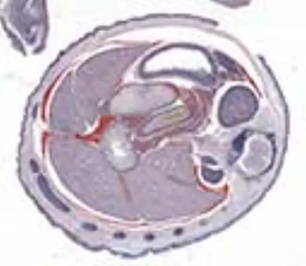
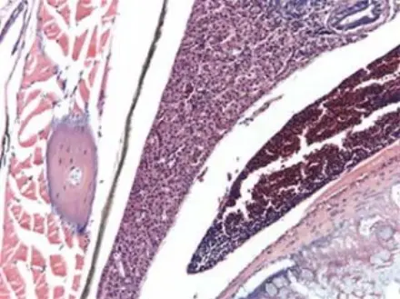
cal1_thumb.jpg

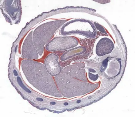
Mouse

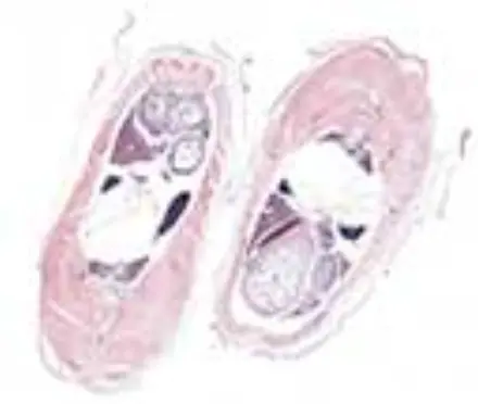
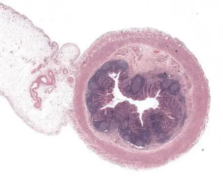
cal2_thumb.jpg

Zebra Fish

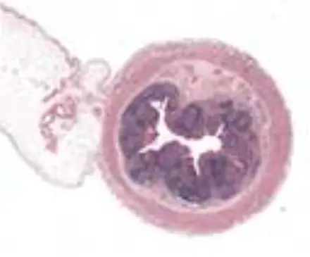
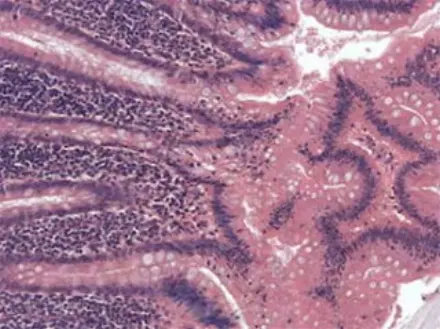
cal3_thumb.jpg

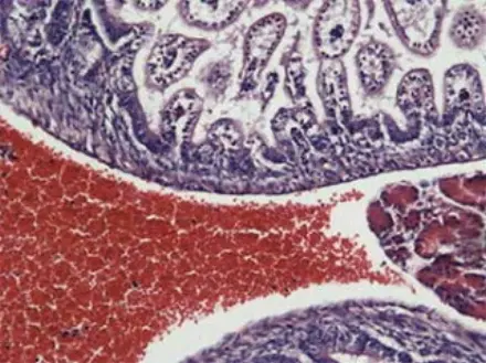
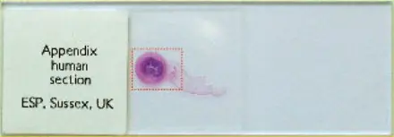
Appendix

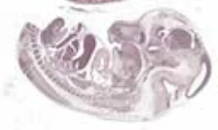
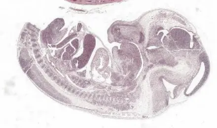
cal4_thumb.jpg

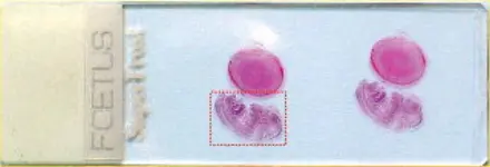
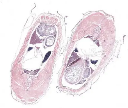
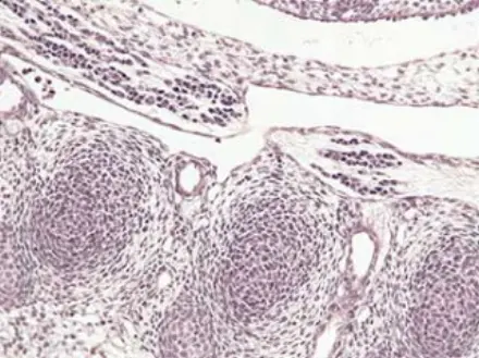
Mouse Foetus

cal4_thumb.jpg

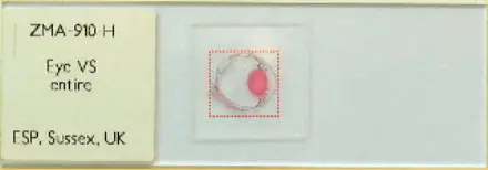
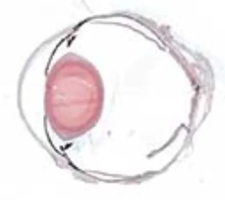
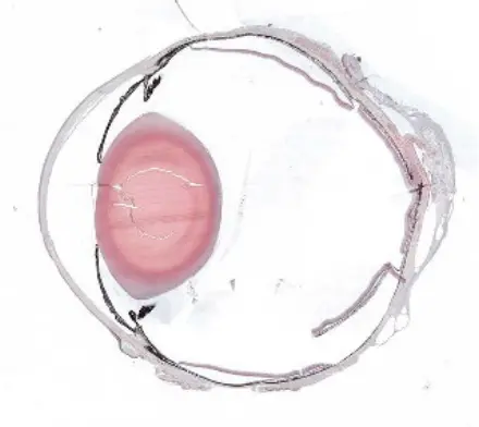
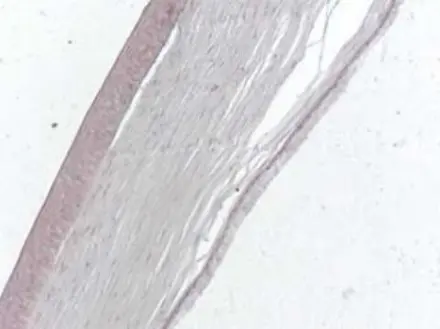
Eye

cal6_thumb.jpg

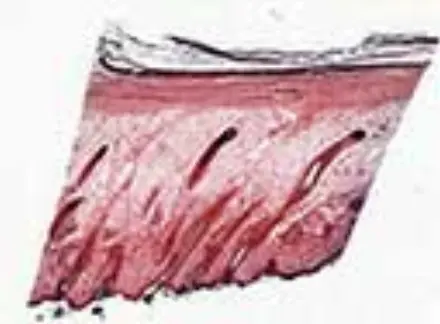
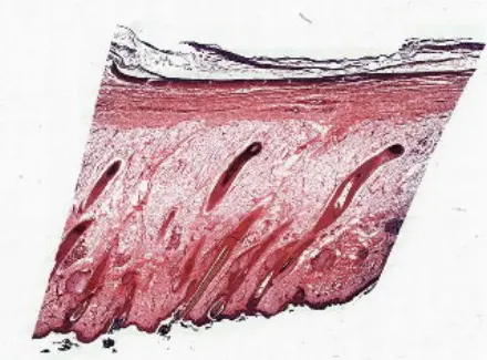
Skin


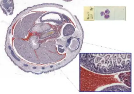
Mouse, Transverse Section

10X Objective / 1X C-mount

Calibration: 0.46 µm/pixel

Scan fields: 17 x 20, 340 total

Size at full resolution: 340 x 4.2 MB = 1.4 GB

Scan area: 10.6 x 9.2 mm = 98 sq. mm

Scan time (snake scan with predictive focus and shading correction): 42 secs

slide1.jpg

掃瞄後影像全覽

cal1_med.jpg

影像放大細節

detailc1_sm.jpg


Zebra Fish

10X Objective / 1X C-mount

Calibration: 0.46 µm/pixel

Scan fields: 16 x 17, 272 total

Size at full resolution: 272 x 4.2 MB = 1.2 GB

Scan area: 9.2 x 7.3 mm = 67 sq. mm

Scan time (snake scan with predictive focus and shading correction): 37 secs

slide2.jpg

掃瞄後影像全覽

影像放大細節

detailc2_sm.jpg


Appendix, Human

10X Objective / 1X C-mount

Calibration: 0.46 µm/pixel

Scan fields: 16 x 18, 288 total

Size at full resolution: 288 x 4.2 MB = 1.25 GB

Scan area: 10.1 x 8.2 mm = 83 sq. mm

Scan time (raster scan with predictive focus and shading correction): 54 secs

slide3.jpg

掃瞄後影像全覽

cal3_med.jpg

影像放大細節

detailc3_sm.jpg


Mouse Foetus, Lateral Section

10X Objective / 1X C-mount

Calibration: 0.46 µm/pixel

Scan fields: 20 x 16, 320 total

Size at full resolution: 320 x 4.2 MB = 1.3 GB

Scan area: 12.5 x 7.4 mm = 93 sq. mm

Scan time (snake scan with predictive focus and shading correction): 46 secs

slide4.jpg

掃瞄後影像全覽

cal4_med.jpg

影像放大細節

detailc4_sm.jpg


Eye Section, Rabbit

10X Objective / 1X C-mount

Calibration: 0.46 µm/pixel

Scan fields: 19 x 23, 437 total

Size at full resolution: 437 x 4.2 MB = 1.8 GB

Scan area: 11.9 x 10.6 mm = 126 sq. mm

Scan time (raster scan with predictive focus and shading correction): 1 min 19 secs

slide5.jpg

掃瞄後影像全覽

cal5_med.jpg

影像放大細節

detailc5_sm.jpg


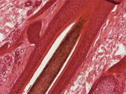
Human Skin, Showing Follicles

10X Objective / 1X C-mount

Calibration: 0.46 µm/pixel

Scan fields: 15 x 15, 225 total

Size at full resolution: 225 x 4.2 MB = 0.95 GB

Scan area: 9.4 x 6.9 mm = 65 sq. mm

Scan time (raster scan with predictive focus and shading correction): 38 secs

slide6.jpg

掃瞄後影像全覽

cal6_med.jpg

影像放大細節

detailc6_sm.jpg


如果您對自動化快速玻片影像掃瞄系統有興趣,歡迎來電洽詢,我們有專業的技術團隊可以提供您滿意的軟體客製化服務。

search_offcanvas

X

Right Click

禁止任意複製或拷貝網站內容!